 Call us:+91-484 2885910 (Reception Orthopedics; 9am to 5pm Mon-Sat), +91-484-2885621 (22,23,24)
Doctor's mobile - +91 9497559755 (whatsApp anytime)
Call us:+91-484 2885910 (Reception Orthopedics; 9am to 5pm Mon-Sat), +91-484-2885621 (22,23,24)
Doctor's mobile - +91 9497559755 (whatsApp anytime)
It is important to assess the ligamentous structures around the knee joint for planning a patellar stabilizing procedure in recurrent dislocation of the patella.
Usually radiographs and MRI scan are done to do the measurement. You will require an expert musculoskeletal radiologist to do accurate measurements to identify alignment issues in the extensor mechanism and trochlear groove of the femur, in addition to Medial patellofemoral ligament tears. Addressing these pathologies, if present, will increase the success rate of a arthroscopic MPFL reconstruction procedure.
Caton Deschamps index
The Caton-Deschamps index is used to measure patellar height and identify patella alta (high riding patella) and patella baja (low lying patella). Patella alta predisposes to patella dislocation.
Normal 0.6 to 1.3
>1.3 – Patella Alta
<0.6 – Patella Baja


Lateral Trochlear Inclination
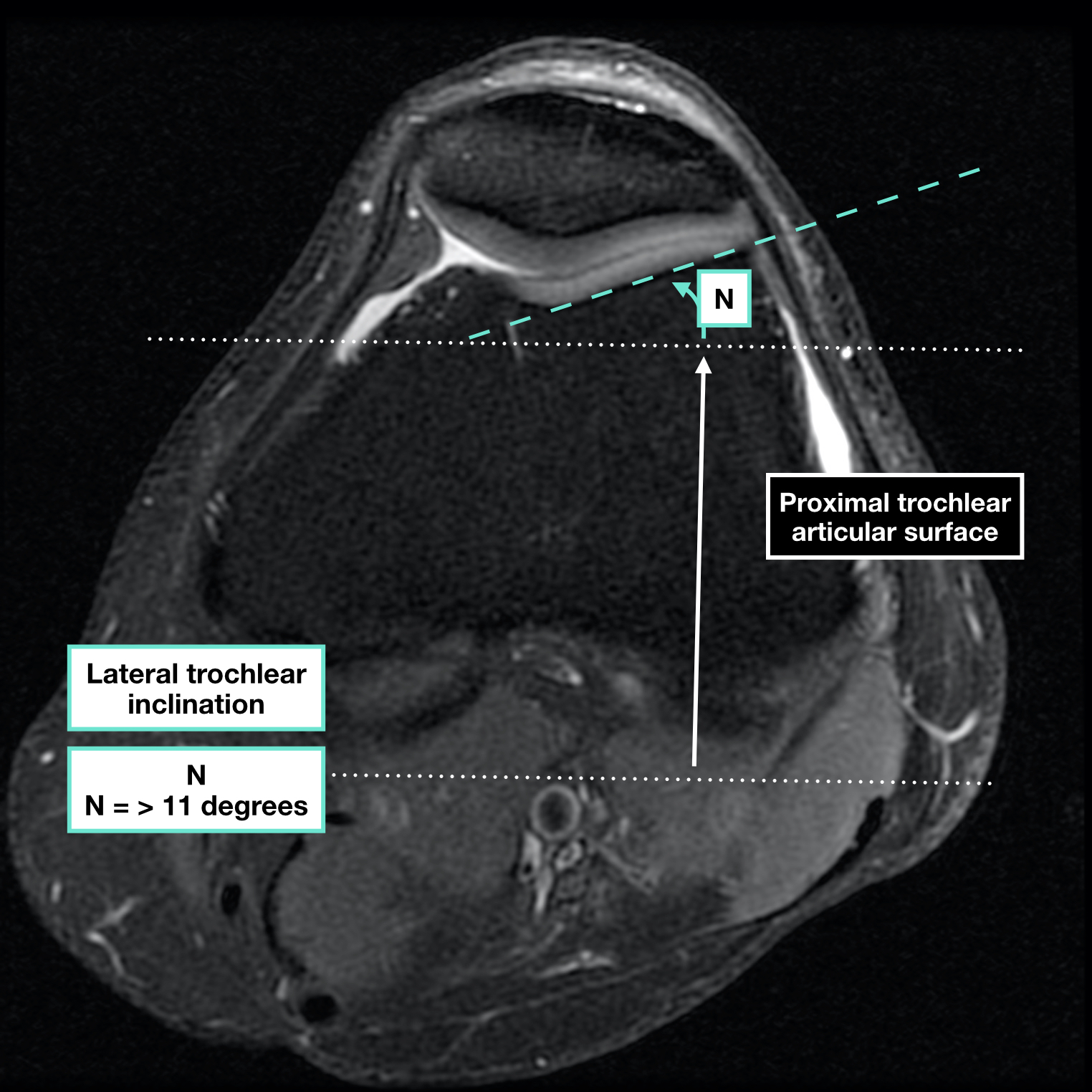
Normal >11 degrees
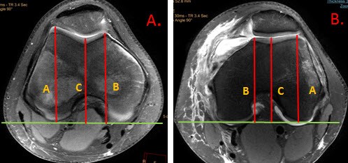
Trochlear facet asymmetry
Normal >0.4


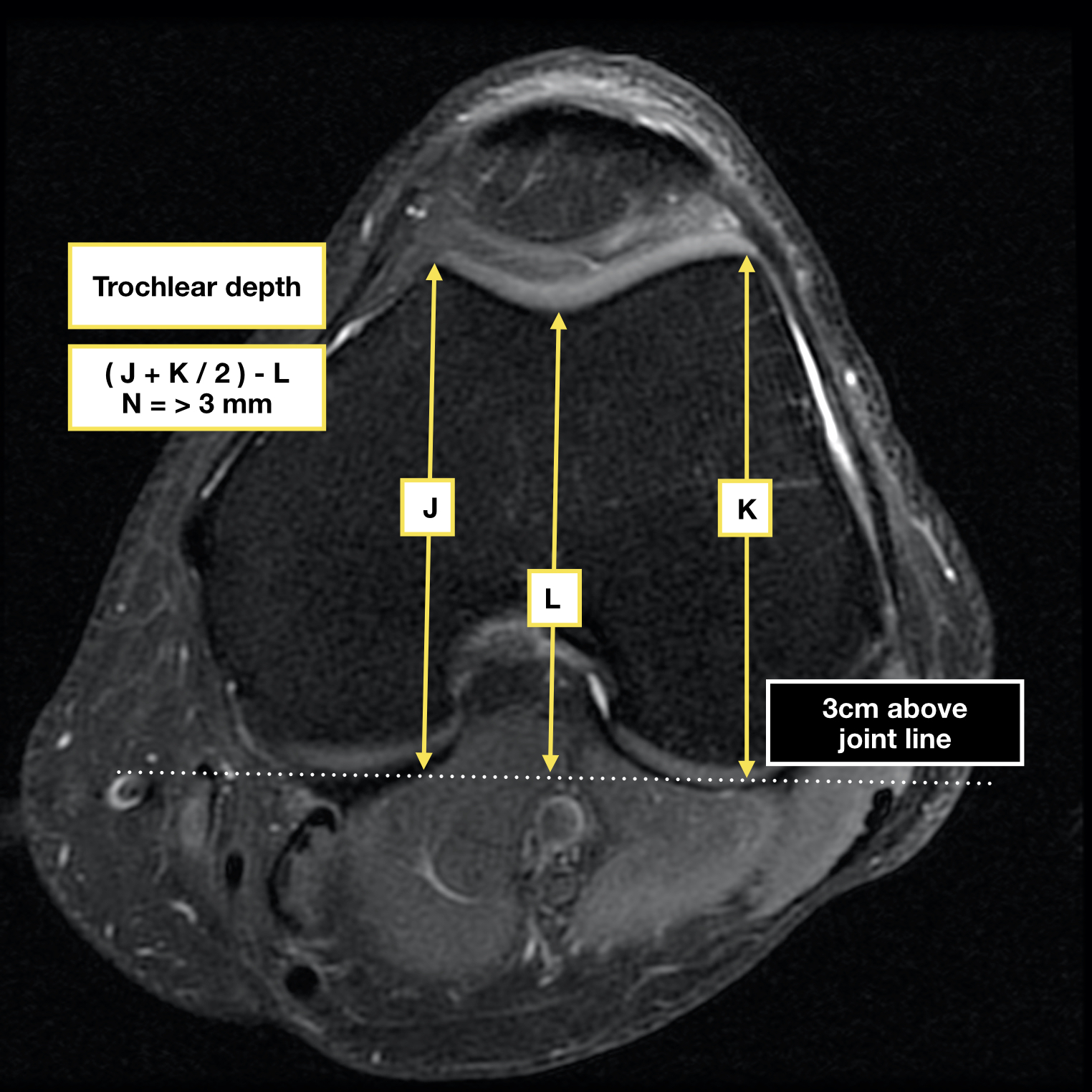
Trochlear depth
Normal >3mm
Trochlear Sulcus Angle (TSA)

Normal 138 +- 6 degrees
TT-TG Distance
Tibial Tuberosity- Trochlear Groove distance
Normal <15mm


TT- PCL distance
Tibial tuberosity – Posterior cruciate Distance
Normal 11.9 +-4.76mm (>20 is abnormal)

Arthroscopic Latarjet changes life of Violinist

Recent Posts

Sr. Consultant Orthopedic Surgeon, Prof & Head,
Institute of Advanced Orthopedics, MOSC Medical College Hospital, Kolenchery, Kochi, Kerala, India - 682311


