 Call us:+91-484 2885910 (Reception Orthopedics; 9am to 5pm Mon-Sat), +91-484-2885621 (22,23,24)
Doctor's mobile - +91 9497559755 (whatsApp anytime)
Call us:+91-484 2885910 (Reception Orthopedics; 9am to 5pm Mon-Sat), +91-484-2885621 (22,23,24)
Doctor's mobile - +91 9497559755 (whatsApp anytime)
Bone loss in shoulder dislocation
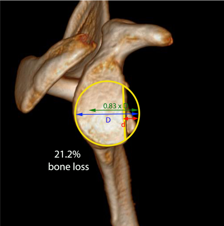
Recurrent dislocation of the shoulder joint leads to bone loss in the glenoid (cup) and humeral head (ball) of the shoulder joint. Treatment of shoulder dislocation is simpler before bone loss develops. Failure to address bone loss can lead to failures in arthroscopic surgeries like bankart procedure. Careful evaluation of bone loss with imaging modalities like CT (3D reconstruction when required) help in determining the most accurate treatment for stabilizing the shoulder joint.
If glenoid bone loss is determined, Dr. Sujit Jos typically recommends surgical stabilization and may offer an arthroscopic glenoid augmentation. Dr. Sujit Jos will utilize bone graft to rebuild bone loss and reconstruct the glenoid during this procedure. Graft options include a Coracoid process (Latarjet Procedure) distal clavicle autograft, iliac crest autograft and distal tibia allograft. In autograft cases, a patient’s own tissue is harvested, either from the coracoid process of the shoudler, clavicle or the iliac crest (largest bone in the hip), and used to reconstruct the area of glenoid bone loss. In distal tibia allograft cases, a portion of a donor’s tibia is harvested and used to perform the reconstruction. Dr. Sujit Jos will explain the appropriate graft option in great detail with each patient at the consultation.
Our special team of Musculoskeletal radiologist will reconstruct the should on a 3D CT scan and measure the bone loss on the glenoid (cup) and humeral head (ball). If there is significant bone loss, a bony augmentation procedure will be adviced by Dr Sujit Jos.
Shoulder dislocation becomes a difficult problem to treat when the glenoid (cup) and humeral head (ball) develops bone loss due to the multiple episodes of dislocation. This page discusses the various options to deal with bone loss associated with shoulder instability.
Read about treatment of Shoulder dislocation before bone loss happens – Arthroscopic Bankart Surgery
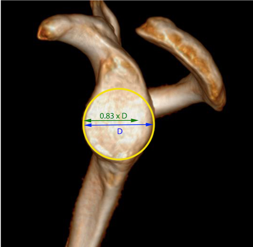
Bone loss assessment
CT scan with 3D reconstruction is done to assess the bone loss in the shoulder bones accurately.


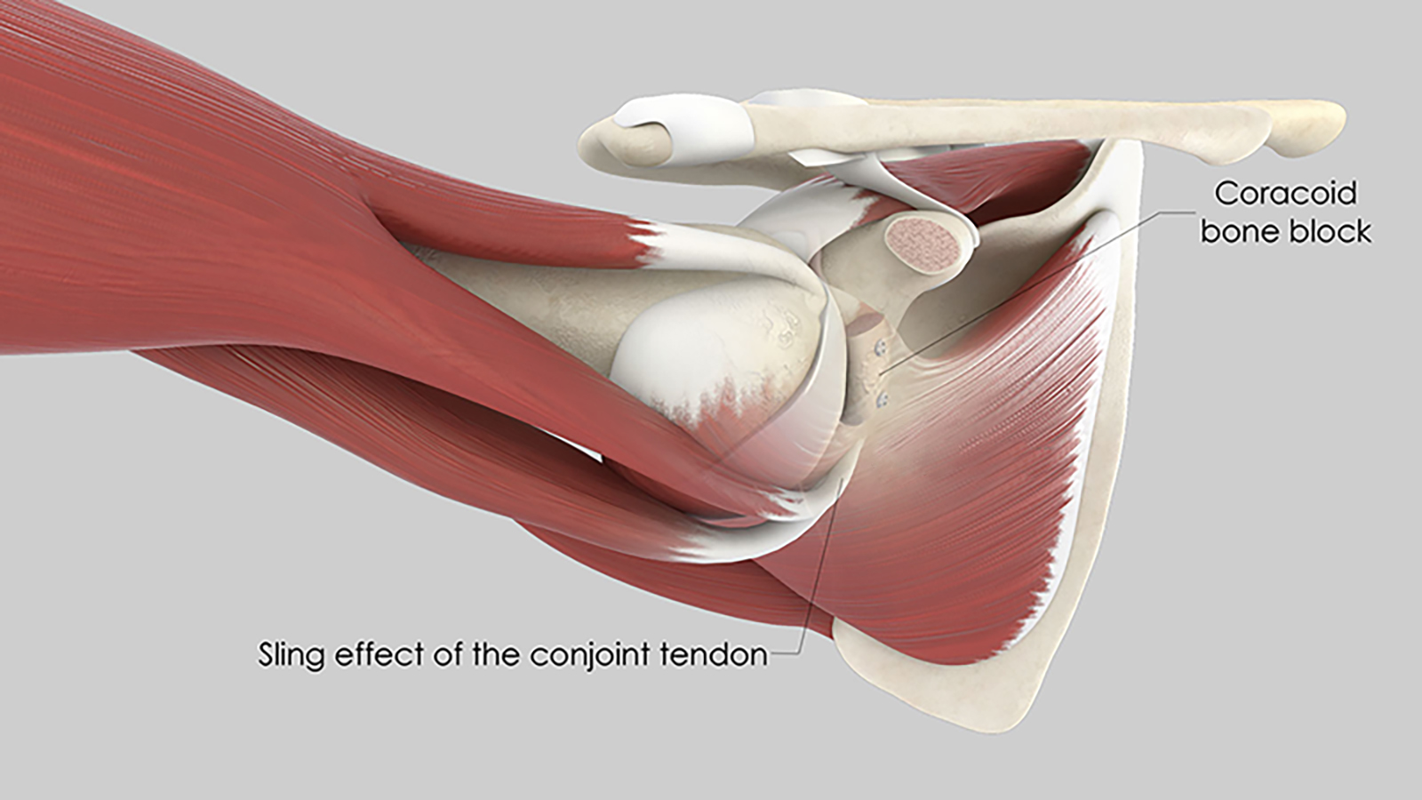
Latarjet Procedure
The common an highly successful bone augmentation procedure for the shoulder joint is an Open or Arthroscopic Latarjet procedure. The Arthroscopic Latarjet procedure has the advantages of less soft tissue injury, faster rehabilitation and ability to correct other joint problems like SLAP tear or a Remplissage for Hill sach lesion during the procedure.

THE OPERATION
BEFORE THE OPERATION
Before the operation, a pre-anaesthesia consultation and a pre-operative assessment are conducted to check the patient is physically apt to undergo the operation and minimise the risk of postoperative complications.
THE OPERATION
The operation takes place in an operating theatre in compliance with strict standards of cleanliness and safety. The patient is placed supine on an operating table. The operation lasts about 2 to 2.5 hours and is carried out under general anaesthesia, which is often combined with an interscalene nerve block. This additional anaesthetic maintains the shoulder and arm numb for several hours and limits postoperative pain.
5 puncture incisions are made in front of the shoulder joint. Stitches will be placed over the incisions to ensure minimal scar. These are removed after 10 days.
AFTER THE OPERATION
The shoulder is numbed with an interscalene block for 12 to 18 hours. Medication and ice also provide effective postoperative pain management.
The surgery can be performed as an in-patient procedurewith a short stay of 2 to 3 days. The patient can return home when discharged.
The shoulder is kept in an orthopaedic sling for 6 weeks. Gentle, passive rehabilitation begins in the week following the operation to stop the shoulder from getting stiff. It is then intensified from the 6th week after the operation and is often completed in the 3rd month after the operation.
When the patient is discharged from the clinic, a consultation with follow-up x-rays is scheduled with the surgeon 4 to 6 weeks after the operation to check the block has not moved and the shoulder is healing well. A second check-up is generally recommended approximately 3 months after the operation.
The duration of medical leaves depends on the patient’s profession but generally varies between 1½ and 2 months. It is longer for manual work.
Driving can be resumed 2 months after the operation.
Sports can be resumed using the shoulder operated on approximately 4 to 6 months after the operation. Usually a CT scan is done after 6 months before resuming sporting activities.
RISKS LINKED TO THE OPERATION
Unfortunately, zero risk does not exist in surgery. Any operation has its risks and limitations, which you must accept or not undergo the operation. However, if an operation is proposed, the surgeon and the anaesthetist consider that the expected benefits far outweigh the risk incurred.
Some risks, such as microbial infections of the surgical site, are common to all types of surgery. Fortunately, this complication is very rare. Bruising can also appear around the surgical site. This is uncommon after Arthroscopic Latarjet procedure and will disappear in few days even if it is seen.
Blood clots can form in the veins in the arm (thrombophlebitis) or lungs (pulmonary embolism). If the anaesthetist considers the risk high, you may be given a treatment to prevent this.
In rare cases, the shoulder remains stiff, hot, and painful for several months after the operation. This complication, known as algodystrophy or Chronic Regional Pain Syndrome (CRPS), is unpredictable and sometimes takes a long time to heal.
Other rarer complications can also occur. Blood vessels (arteries, veins) can be accidentally damaged and will require vascular surgery (bypass). Nerves can also be damaged accidentally during the operation with a risk of paralysis or loss of feeling in the limb operated on, which can be transitory or permanent. But these are fortunately very rare in the hands of an expert surgeon like Dr Sujit Jos.
Finally, the bone block may not heal and may be torn off with time and the strain.
If you have any concerns about the operation, do not hesitate to talk to your surgeon or the anaesthetist and they will answer any questions you may have.
ADVANTAGES OF ARTHROSCOPIC LATARJET PROCEDURE
Several advantages are linked to Arthroscopic Latarjet procedure, although it takes a bit longer than an open traditional Latarjet procedure. The main advanatage is that arthroscopic visualizaiton allows accurate placement of the coracoid graft in its position in front of the glenoid where there is bone loss. The scarring will be minimal and will almost disappear in few months time. Unlike in open latarjet, associated pathology like a SLAP tear, labral tear and an off-track Hill sachs lesion can be effectively treated in Arthroscopic method of Latarjet procedure. Another major advantage is that the labrum can be reattached to its position in front of the glenoid in Arthroscopic Latarjet procedure. Dr Sujit Jos is the pioneer surgeon to do Arthroscopic Latarjet procedure in South India and is a trainer surgeon for this procedure in India.
Understand Arthroscopic Latarjet procedure with Titanium cancellous screws
Arthroscopic Latarjet procedure with All-fibre method (Metal Free)
Advantages of Metal free Arthroscopic Latarjet procedure
- Less soft tissue dissection
- Reduced pain
- Faster recovery
- Safe from head of humerus impingement on the metal screws as in standard Latarjet procedure 5. Minimal scars – better cosmesis
- Dynamic sling effect of the conjoint tendon as in Latarjet transfer
- As it is an arthroscopic technique, anterior capsule repair can be done concomitantly providing a smooth surface for the movement of the humeral head, thereby protecting from wear Although the learning curve is steep, this technique is being embraced by Shoulder surgeons all around the world for treating shoulder dislocation when bone loss has set in after many episodes of instability of the shoulder.

Movement of the shoulder 6 weeks after Arthroscopic Latarjet procedure
Useful Links:
https://www.ncbi.nlm.nih.gov/pmc/articles/PMC4437927/
http://www.clevelandshoulder.com/articles/arthro-latarjet.pdf
Arthroscopic Latarjet changes life of Violinist

Recent Posts

Sr. Consultant Orthopedic Surgeon, Prof & Head,
Institute of Advanced Orthopedics, MOSC Medical College Hospital, Kolenchery, Kochi, Kerala, India - 682311


